

Tissue-Mimicking Materials
Project Overview
The last few years have seen a dramatic rise in AM in the medical field. Many medical facilities are turning to printed models taken from CT scans to better visualize complex anatomies prior to a procedure. These models can also be used for surgical planning, education, training, and communication with patients/families. In order to create realistic structural models, materials must be carefully selected to closely match the behavior of specific cardiac tissues.
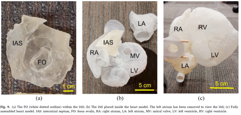
By using material jetting to explore the combination of a rubber-like photocuring material and a non-curing liquid, a material mimicking the material properties of the fossa ovalis was determined. In collaboration with cardiologists at the Carilion Research Institute in Roanoke, a full-scale heart model was fabricated integrating the tissue-mimicking material. Cardiologists confirmed the visual and structural realism of the fossa ovalis within the printed heart model. The model will further be used for medical student trainees to practice the transseptal puncture procedure.
Future medical models with tailored material properties will enable more realistic surgical simulations for a variety of procedures.
Related Publications
L. Bezek, M. Cauchi, R. De Vita, J. Foerst, and C. Williams, “3D Printing Tissue-Mimicking Materials for Realistic Transseptal Puncture Models,” Journal of the Mechanical Behavior of Biomedical Materials, Vol. 110 103971 (2020)